
A bruise can outlast your memory of the bump that made it. Yesterday's dark patch may acquire a yellow edge, while another part stays purple. It looks almost as though the skin is trying out colours. In fact, you are watching escaped blood being dismantled and removed, through a covering that changes how those colours appear.
The chemistry has an order. The visible colours do not provide a dependable timetable. Understanding the difference explains both the changing patch and why a photograph cannot reliably tell you when an injury happened.
Blood in the wrong place
A knock can damage small blood vessels without opening the skin above them. Blood escapes into the surrounding tissue. Red blood cells that normally travel inside vessels are now outside their usual route, and their coloured contents contribute to the mark. MedlinePlus describes bleeding beneath the skin.
The body must stop further leakage as well as deal with blood already lost into tissue. Tiny blood fragments called platelets gather at vessel damage, and a network of the protein fibrin reinforces the plug. Sealing the leak does not remove the escaped cells. The NIH explains the clotting response.
Inside each red blood cell is haemoglobin, the oxygen-carrying protein that gives blood its colour. Its light absorption changes with how much oxygen it carries and with its chemical state. Human bruise measurements track changes in blood content and oxygenation. But the patch you see is not a sample of blood held up in a clear glass.
Skin changes the view
Light must enter skin, interact with tissue and pigments, and return to your eyes. Some colours of light are absorbed more strongly than others; tissue also scatters light. The blood's depth, the skin above it and the lighting therefore affect the visible colour. Blue or purple does not mean that the body has manufactured blue blood. Optical researchers explain why bruise depth matters.
Imagine two otherwise similar patches of leaked blood, one shallow and one deeper. Even with the same amount of pigment, they need not look alike because the light takes different paths through tissue. Now change the room's lighting without changing the bruise. Its appearance can change again. This is an illustrative comparison, not a formula for estimating depth or age.
Skin tone matters too. The pigment melanin affects the contrast between bruised and unbruised tissue. A 2026 review found that bruises are harder to detect visually on darker skin, with important gaps remaining in real-world evaluation of imaging methods. Reduced visibility is not evidence of less injury. The review maps these detection limits.
The cleanup changes the molecules
Immune cells called macrophages engulf escaped red blood cells and help break down their contents. Haemoglobin contains an iron-bearing part called heme. Dismantling heme produces different molecules, with different light-absorbing properties, rather than simply diluting the original red colour.
Proteins called enzymes help chemical reactions happen. One enzyme, heme oxygenase, opens heme's ring structure. This produces biliverdin, a green pigment, and releases iron. Another enzyme quickly converts biliverdin into bilirubin, a yellow-orange pigment. The biochemical pathway separates these products.
In a rat bruise study published in 2015, researchers found strong heme oxygenase-1 expression in macrophages. The finding supports a role for recruited cleanup cells in pigment formation. It does not supply a clock for human bruises. The original study describes its animal model.
There is a tempting shortcut here: red blood, then green biliverdin, then yellow bilirubin, each assigned a day. But a chemical intermediate need not accumulate enough to become a visible stage. In a 2004 human study, researchers found no distinct light-absorption peak indicating biliverdin accumulation, consistent with its rapid conversion. Their measurements linked yellowing to local haemoglobin breakdown. Read the spectrophotometry study.
Nor does an apparent green necessarily identify one green chemical. Haemoglobin and bilirubin absorb overlapping parts of the spectrum, so their combined appearance can be mistaken for biliverdin. Researchers explicitly warn against this identification.
Where the colour goes
Conversion is not the same as removal. A yellow pigment still contributes colour while it remains in the tissue. As material is transported away, the amount of pigment left locally falls. Bilirubin entering the bloodstream travels bound to albumin, a carrier protein. The liver changes it into a form that can be secreted in bile and delivered to the intestine. A physiological review follows that transport route.
Iron takes a different route. Cells can store it and make it available for reuse. Some local iron-containing material forms haemosiderin, a brownish pigment that can contribute to residual discoloration. It is not bilirubin turning brown, and not every bruise develops an obvious brown stage. Macrophage research describes iron storage and recycling; the bruise study discusses haemosiderin.
The result is a changing mixture. Different parts of a bruise can contain different amounts of blood and breakdown products. A yellow margin around a darker centre is therefore compatible with processes occurring together, not evidence that the margin was injured on a different date.
Think of the process as four overlapping jobs: blood leaks out, red cells remain in tissue, cleanup cells dismantle their pigments, and breakdown products are cleared or recycled. These are changes in cells, molecules and location, not four colours that every bruise must display.
Blood leaves a vessel; its pigments are changed and cleared
Overlapping processes, not a colour timeline
Blood leaks into tissue
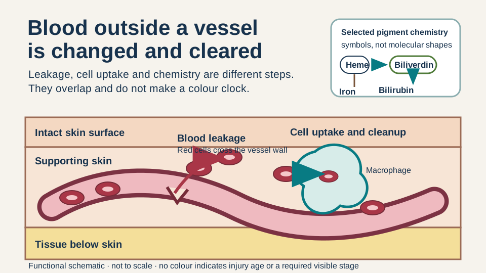
Damaged vessel. Red cells leave vessel.
Skin surface stays closed.
Cells remain outside the vessel
Leak sealed. Cells remain in tissue.
Haemoglobin inside a red cell.
Cleanup changes pigments
Cell uptake. Macrophage takes up cells.
Haemoglobin is broken down; heme is released
Chemical insetSymbols are not molecular shapes · Selected products onlyHemeIron released↓Heme oxygenaseBiliverdin↓Rapid chemical conversionBilirubinMaterial leaves or is retained for reuse
Pigment clearance from tissue follows the dashed arrow.
Iron stored / recycled; some haemosiderin remains.
Systemic route; simplifiedBilirubin: blood carrier → liver → bile
Each panel keeps the skin surface, tissue layers and small vessel in the same positions. First, red blood cells move through a break in the vessel wall into tissue. Second, the leak is sealed but red cells remain outside; their haemoglobin is still inside the cells. Third, a macrophage takes up a red cell. A separate chemical inset shows heme changing downward to biliverdin and then bilirubin, with iron released along a branch from heme. Fourth, a clearance arrow points toward the edge of the local tissue field, while a labelled cell retains some iron-containing material. A separate text route follows bilirubin through blood transport, liver processing and bile. The numbered jobs can overlap and do not specify the bruise's visible colour or age.
Schematic, not to scale. Cells, molecules and residue amounts are symbolic. Processes overlap; panel spacing and colours show neither elapsed time nor the expected appearance on any skin tone. Not an injury-age or safety test.
Sources: Langlois et al. (2015): macrophage enzyme evidence in a rat model; Hughes et al. (2004): local pigments; Stam et al. (2012): transport and spatial limits; Kipp et al. (2025): systemic bilirubin handling. The selected pathway omits amino-acid recovery, other scavenging routes, detailed lymphatic anatomy, carbon monoxide and biochemical reactants.
Why a colour chart cannot date the bump
Forensic researchers have tested the appealing idea that colour reveals age. In 1991, investigators examined 369 photographs from 89 people. Yellow was the most informative colour change, but its development varied with age, and red did not change significantly with time. Those findings were much narrower than a universal colour calendar. The study reports the observations and their limits.
A 2009 review concluded that colour should not be the main basis for dating a bruise, describing the available evidence as limited and inconsistent. Measuring pigment chemistry with instruments is a different undertaking from matching a photograph to a chart. Neither the existence of chemical changes nor a promising research method validates an everyday visual clock. The forensic review explains the distinction.
Why a colour chart cannot be a clock
Colour is not a safety test
This is an explanation of ordinary bruising, not a diagnosis. Unexplained or unusually easy bruising deserves medical advice, especially with other bleeding. Do not change prescribed medicines on the strength of a bruise's colour. Merck's clinical guidance explains when assessment is needed.
After an injury, severe or worsening pain, increasing swelling or difficulty using the limb needs urgent assessment. Numbness, deformity or a cold injured limb needs immediate care. A yellow edge does not cancel these warnings. The NHS distinguishes urgent and emergency signs.
A fading bruise is not a calendar printed beneath the skin. It is the visible trace of a cleanup: blood components changing form, useful material being recovered, and colour-bearing molecules leaving the scene.
YOUR PERSPECTIVE
Join the discussion
What surprised you? Have a question or a correction? Share your thinking.
No comments yet. Start the conversation.
Sign in with ChatGPT to commentAnyone can read the discussion. Sign-in helps limit spam.