
A small cut can stop producing fresh blood while the split in the skin is still easy to see. The body has solved one urgent problem: blood escaping through damaged vessels. It has not yet finished replacing the damaged tissue. Stopping bleeding is called haemostasis. It is an early part of healing, not proof that healing is complete.
Think of sealing a leak before rebuilding the damaged wall around it. That distinction helps explain the cut, although the body has no separate emergency crew that packs up before the builders arrive. Its responses overlap. Even the temporary seal helps organise the repair. Injured vessels can narrow to reduce blood loss, while blood components assemble a plug at the damage. NHLBI: how blood clots
How the plug finds the damage
Blood already carries tiny cell fragments called platelets. When a vessel is damaged, material beneath its inner lining becomes exposed. That material includes collagen, a structural protein. The injury therefore presents attachment sites that passing platelets would not normally meet.
A protein called von Willebrand factor helps platelets catch onto the exposed surface, especially where flowing blood would otherwise sweep them past. Platelets also have attachment machinery that recognises collagen directly. These contacts help switch platelets into an active state.
Activated platelets change shape and release chemical signals that activate nearby platelets. Their surface proteins can bind bridging molecules, including the soluble blood protein fibrinogen, so neighbours join together. A local cluster grows into a plug. This is more specific than blood simply becoming sticky: particular molecules connect the injury, the first platelets and the platelets arriving after them. The details vary with the injury and the forces of flowing blood. Review: von Willebrand factor and platelets
A loose cluster gains reinforcement
While platelets gather, linked chemical reactions generate thrombin, an enzyme that cuts particular proteins. Thrombin cuts a soluble blood protein, fibrinogen, producing building blocks of fibrin. These blocks join into fibres. Additional chemical links strengthen the resulting network.
The fibrin network reinforces the platelet plug. Imagine gathering loose buttons inside a net: connecting the material changes how well it holds together. The analogy stops at the chemistry. Fibrin is made locally from material carried in the blood, and the platelets actively participate in clot formation. They are not passive objects dropped into a ready-made bag.
Platelet activation and fibrin formation support each other. Thrombin also activates platelets, and activated platelet surfaces help organise clotting reactions. The process does not wait for a finished platelet plug before starting its reinforcement. Review: current understanding of haemostasis
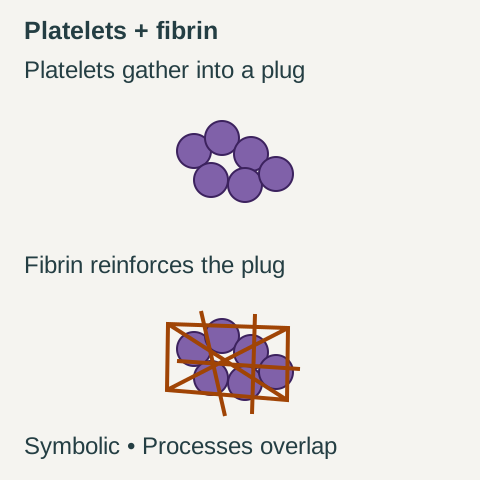
A platelet plug gains a fibrin network
Platelets gather
Purple discs: platelets. These cell fragments stick at the damaged site and join one another.
Fibrin adds reinforcement
Orange lines: fibrin fibres. A protein network forms through and around the gathered platelets.
Symbolic comparison, not to scale or a cutaway. Platelet activity and the reactions that make fibrin overlap. The panels omit other blood cells, vessel walls and most clotting reactions; they show neither a healing timetable nor a clinical prediction.
Original diagram by The Daily Wonder. Sources: NHLBI: How blood clots; Weisel and Litvinov (2017), fibrin formation.
The seal can pull itself tighter
Platelets can grip fibrin fibres and pull on them using contractile machinery inside the platelet. Many such pulls compact the network. This is clot contraction, also called clot retraction. It can make the clot denser and bring the wound edges closer together.
That movement is useful because an effective seal needs more than an accumulation of material: its parts must resist coming apart. The blood clot is a structure being assembled and mechanically adjusted. The red blood cells caught within it help explain why a visible clot looks red, but platelets and fibrin provide crucial organising and supporting roles. Contracting this temporary structure still does not rebuild the layers of skin. Review: the mechanisms of clot retraction
Repair means making living tissue
The clot also provides a temporary framework through which repair cells can move. Immune cells help deal with microbes and remove damaged material. This inflammatory response overlaps with the work of rebuilding the wound; it is not a separate job that must always finish first.
Cells responsible for the skin's outer covering move and multiply to restore a continuous surface. Where deeper support tissue has been damaged, cells called fibroblasts produce replacement material, including collagen. New small blood vessels can supply the developing tissue. Later, the repair tissue is reorganised through remodelling.
These tasks explain why stopping visible bleeding and restoring skin take different amounts of work. A seal can interrupt a route for blood to escape without replacing every damaged cell or restoring the tissue's strength. If a scar forms, it consists of repaired tissue with a changed structure. It is not simply the original blood clot left permanently in place. Review: wound healing from a cellular perspective
A scab is a surface crust
The familiar scab is related to clotting, but the words are not interchangeable. A surface layer containing fibrin and dead cells can dry into a crust. The clotting response also operates at injured vessels, rather than being only the visible crust on top of the skin.
Living skin cells restore the covering beneath or around the wound's surface material. Forming a dry scab is not a necessary biological checkpoint before skin can close; repair can also proceed in a moist environment. Nor does a scab's appearance tell us that the deeper tissue has finished repairing. The surface and the tissue below it are doing different things. Review: cellular mechanisms of skin repair
The temporary network must be removed
A successful repair must also deal with the material it no longer needs. The body has a regulated system for breaking down fibrin, called fibrinolysis. An enzyme called plasmin cuts the fibres. Other molecules control plasmin's production and activity, so removal depends on a balance of opposing processes.
This is active dismantling, not simply blood drying up. The structure of a clot affects how easily enzymes can get into it and break it down. Formation, contraction and removal therefore influence one another. There is no single clock that makes every minor cut pass through neat, non-overlapping stages. Removing fibrin and rebuilding skin are coordinated parts of recovery, rather than one universal final switch. Review: how fibrinolysis works
Seeing the workers in the blood
In studies reported in 1881–82, the Italian researcher Giulio Bizzozero used microscopy of circulating blood and experiments with blood flow to establish platelets' role in stopping bleeding and forming clots. Other observers had probably seen platelets earlier. His contribution was to connect these small structures with what they did. That history captures the key insight: a clot is an organised response involving active components of blood. The cut stops bleeding because it gains a temporary seal; healing continues because living tissue still has to be repaired. Historical review: platelet research
Why a “clotting cascade” is only a map
YOUR PERSPECTIVE
Join the discussion
What surprised you? Have a question or a correction? Share your thinking.
No comments yet. Start the conversation.
Sign in with ChatGPT to commentAnyone can read the discussion. Sign-in helps limit spam.