
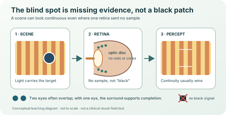
A damaged camera sensor often announces itself with a dead pixel. The human eye has something much larger: a patch in every retina with no rods or cones, the cells that respond to light. Yet ordinary vision does not contain a dark oval floating beside whatever you look at.
That sounds as if the brain performs a clever repair. But the first important clue is simpler: missing input is not the same thing as black input. A black patch would be a positive report that a region is dark. At the blind spot, no photoreceptor makes any report at all.
Vision therefore faces a gap in evidence, not a black object. Most of the time it resolves that gap so quietly that the gap never becomes an object of experience.
The exit that cannot also be a sensor
The retina is a sheet of nervous tissue at the back of the eye. Rods and cones convert light into electrical signals. Several layers of retinal cells process those signals, and retinal ganglion cells send the result onward through long fibres called axons.
Those axons must leave the eye together. They converge at the optic disc, pass through the back of the eyeball and become the optic nerve. Blood vessels also enter and leave there. The price of that exit is a patch without photoreceptors. The corresponding region of the visual field is the physiological blind spot. The US National Library of Medicine’s neuroscience text describes the optic disc as the retinal exit point that contains no photoreceptors.
The blind spot lies off to the side rather than at the centre of gaze. Its border varies among people and is not perfectly crisp. High-resolution measurements place it roughly 15 to 20 degrees into the temporal, or outward, field. The 2023 fine-scale mapping study is evidence from measured eyes, not a universal template.
Two eyes make the first gap disappear
Your two blind spots fall on opposite sides of the combined visual field. For much of the scene, a location that lands on one eye’s optic disc is still sampled by the other eye. The brain can use that available evidence when both eyes are open.
But binocular overlap cannot be the whole explanation. Close one eye and the world still does not gain a black oval. The visual system has to deal with the missing input from the remaining eye.
The brain completes relationships, not hidden facts
What appears in the monocular blind spot depends on what surrounds it. A uniform field tends to look uniform. A line that reaches both sides of the gap can appear continuous. Some textures and colours also extend perceptually across it. Researchers call this perceptual filling-in: a visual attribute is experienced inside a region even though the relevant attribute exists only around that region. A peer-reviewed taxonomy of filling-in shows why the term covers several distinct phenomena rather than one universal paintbrush.
“Filling-in” can suggest that the brain copies real scenery into a hole. It does not. The system can continue a colour, texture or contour when the surroundings support it, but it cannot recover an unseen word or identify a hidden face from absent data.
The result is a constrained best fit. Vision already groups edges, surfaces and objects across noisy or partly blocked scenes. At the blind spot, those rules meet a region where direct evidence has always been unavailable. Matching surroundings favour continuity; conflicting surroundings can produce different percepts.
To notice a hole, vision would need evidence for its boundary. The optic disc supplies no black border and no “data missing” label. It supplies silence.
Mariotte turned silence into an observation
In the seventeenth century, the French experimenter Edme Mariotte showed that an object could vanish at a particular position while another point was fixated. His 1668 book, Nouvelle découverte touchant la veüe, survives in a digitized copy from the Getty Research Institute. The demonstration mattered because the optic-nerve entrance had been treated as a privileged part of the eye. A repeatable failure of vision argued otherwise.
The demonstration revealed the blind spot indirectly: a chosen target disappeared while the background remained perceptually present. That distinction remains central to modern experiments.
What the model can and cannot show
EXPLORE THE IDEA
When does a target land on the optic disc?
Point eye and screen
Top-down schematic: upward on this page represents sideways offset from fixation on the screen, not height in the visual field.
14.6°Target angle
Inside12–18° band
AvailableFellow-eye evidence
- Point eye: outlined circle on the left. The vertical line on the right is the screen.
- Fixation: cross on the screen, joined to the eye by the dashed line.
- Target: filled orange dot on the screen, joined to the eye by the solid teal ray.
- Selected angle: curved ink mark between the dashed fixation line and target ray, currently 14.6°.
- Dimensions: bottom bar = 50 cm viewing distance; upright bar beside the screen = 13 cm sideways offset. Both axes use the same fixed scale.
- Illustrative 12–18° band: gold fan near the point eye. This angular comparison is not a retinal location, anatomical border or personal blind-spot measurement.
Evidence ledger for this illustrative model
This eyeno target sample
Fellow eyeevidence available
Surroundingscontext remains
Keep the target offset fixed and increase viewing distance. Its visual angle shrinks, so it can move out of the illustrative blind-spot band even though the marks on the screen did not move.
This is an illustrative geometry model, not a test of eyesight. It treats the eye as a point and uses a fixed 12–18° band only to show the idea; real blind-spot position and borders vary, and eye fixation must be controlled. Fellow-eye coverage is simplified to on or off. The model does not simulate retinal optics, neural filling-in or clinical visual fields, and it cannot diagnose a vision problem. No animation.
Neurons respond when nothing was sampled there
Filling-in is not merely a philosopher’s label for our ignorance. Experiments can ask whether brain activity changes when a stimulus arrangement produces a filled-in percept.
In macaques, Hidehiko Komatsu and colleagues recorded neurons in primary visual cortex, or V1, whose mapped visual locations lay inside the blind-spot representation. Some neurons responded to large, uniform surfaces covering the blind spot. The original 2000 study supports an active neural response associated with completion. It does not prove that one patch of cortex literally contains a finished miniature picture.
Human brain imaging has since mapped the cortical territory around the blind spot in finer detail. A 2022 retinotopic study also found evidence consistent with completion beyond V1. Scientists still debate the computations, timing and brain areas behind different kinds of filling-in.
The blind spot has even become an experimental tool. Researchers can arrange for a target change to remain physically invisible until an eye movement shifts the blind spot away from it. A 2021 methods paper uses this property to study perception across rapid eye movements without pretending that the blind spot is an empty black mask.
A seam hidden by the way seeing works
The blind spot is often presented as a party trick: make a dot disappear. Its deeper lesson is about what a visual experience is.
The eye does not transmit a complete photograph, and the brain does not display missing pixels as an error message. Two eyes provide overlapping evidence. With one eye, surrounding structure usually supports continuity.
That makes the seamless scene useful, but not infallible. Perception is an evidence-based construction whose edits are normally invisible to its owner. The small hole in the retina disappears not because the brain knows what was there, but because it can often decide what kind of relationship should continue through a place where nothing was sampled.
YOUR PERSPECTIVE
Join the discussion
What surprised you? Have a question or a correction? Share your thinking.
No comments yet. Start the conversation.
Sign in with ChatGPT to commentAnyone can read the discussion. Sign-in helps limit spam.