
A kidney performs an apparent contradiction. It lets an enormous stream of water and small dissolved substances leave the blood, then prevents most of that water from leaving the body.
The contradiction disappears when filtration and excretion are separated. The kidneys process roughly 120–150 quarts of blood each day, while the urinary tract normally carries away only about 1–2 quarts of urine. Those figures, from the US National Institute of Diabetes and Digestive and Kidney Diseases, are not a claim that the body owns that much blood. The same circulating blood repeatedly passes through the organs, and useful water and solutes are reclaimed from the filtered fluid.
The most surprising part of that recovery is not a stronger filter. It is a two-stage information system written into the kidney's anatomy: first build a chemical slope; then decide whether the final stream of fluid is allowed to feel it.
A straight pipe would waste the difference
Each kidney contains many microscopic processing units called nephrons. Fluid filtered from a tuft of capillaries enters a tubule. Much of its water and useful material is recovered early, but the final ability to conserve water depends on a region called the medulla, beneath the outer cortex.
A straight tube crossing this region could exchange water and salt with its surroundings, but it would not easily create a large, orderly difference from top to bottom. The nephron instead folds into a hairpin called the loop of Henle. Fluid goes down one limb and returns in the opposite direction through the other.
That opposing flow is the geometric trick. The descending limb readily loses water. The ascending limb is extremely resistant to water movement, while salt can leave it; in the thick ascending limb, transport proteins actively move sodium and chloride out. A small side-to-side separation at each level is repeated along the length of the loop. The result is a vertical gradient: the tissue becomes progressively more concentrated deeper in the medulla.
Tubules build the difference; vessels limit its loss
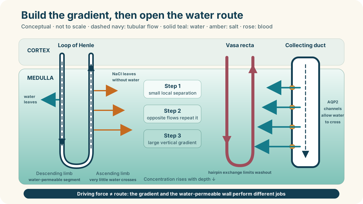
The top band represents the cortex. Below it, the medullary tissue becomes more concentrated with depth. These panels separate the loop’s building role from the vessels’ preserving role
Loop of Henle: fluid flows down, then up
Descending limb: water can leave. Teal arrows point from a water-permeable segment into the surrounding tissue
Ascending limb: NaCl can leave; very little water crosses. Amber arrows show salt transfer. In its thick segment, transport proteins spend energy to move salt out
Vasa recta: limit gradient washout
Blood also flows down, then returns up. Descending blood loses water and gains solutes; ascending blood exchanges in the reverse directions
The hairpin exchange helps retain medullary solutes while the vessels carry recovered water away. It limits washout; it does not make the gradient permanent
Dashed navy arrows inside the loop: tubular-fluid direction. Dashed rose arrows inside vessels: blood direction. Solid teal arrows across walls: water exchange. Dotted amber arrows: NaCl leaving the loop, or solute exchange at vessels. Darker green bands: more concentrated tissue at depth
Conceptual teaching sections, not to scale or a complete nephron. The loop panel simplifies the established outer-medullary mechanism: the thick ascending limb actively transports NaCl. Descending segments vary in water permeability. Real vessels form networks; blood flow affects washout. Bands show direction of increasing concentration, not measured steps or osmolality. Urea and three-dimensional structure matter, and the complete inner-medullary mechanism remains unsettled. Not diagnostic or guidance for drinking water.
Sources: Gottschalk and Mylle: original countercurrent evidence; Sands and Layton: outer-medullary mechanism and inner-medullary limits; Pannabecker and colleagues: medullary architecture; Warshaw and colleagues: new inner-medullary architecture evidence (2026)
Physical chemist Werner Kuhn and Kaspar Ryffel proposed this countercurrent multiplication principle in 1942. It took years to gain experimental support. In 1959, Carl Gottschalk and Margaret Mylle sampled fluid from tiny tubules in living animal kidneys. Their micropuncture measurements provided evidence for the countercurrent model; the American Physiological Society preserves the study and its history.
The staircase is stored in the tissue
“Salt staircase” is a picture, not a row of crystals. The steps are increasing osmolality: the number of dissolved particles per kilogram of water. Near the cortex, the surroundings are roughly similar in concentration to blood plasma. Farther down the medulla, sodium chloride and urea make the surroundings more concentrated.
The loop does not concentrate the final urine by itself. It builds the surroundings that can draw water later. That distinction is easy to miss. A pump can create the gradient, but another structure must expose the outgoing fluid to it.
The architecture is also more intricate than the familiar single hairpin in a textbook. Loops of different lengths, collecting ducts and bundles of blood vessels occupy particular three-dimensional arrangements. Modern anatomical work shows why the relationships among tubules and vessels matter to the concentrating mechanism.
Blood must collect water without erasing the staircase
Reabsorbed water has to return to the circulation. Yet ordinary blood flow through the medulla could carry away its accumulated salt and flatten the gradient—a problem physiologists call washout.
The solution is another hairpin system: the vasa recta, narrow blood vessels that descend into the medulla and climb back out. As blood descends, it exchanges water and solutes with increasingly concentrated tissue; as it ascends, the exchange reverses. This countercurrent exchange lets the vessels carry recovered water away while limiting the loss of the medullary gradient.
“Limiting” matters. The gradient is dynamic, not painted permanently into the kidney. Blood flow, salt transport, urea movement and water flow all influence it. Detailed reviews describe the medulla as an organised network rather than two isolated pipes; three-dimensional studies have tested how that organisation supports the gradient.
The final tube receives a permission signal
Filtered fluid eventually enters a collecting duct, which runs from the cortex through the medulla. Now the kidney can spend the gradient it built.
When the body needs to conserve water, the hormone vasopressin, also called antidiuretic hormone, binds to receptors on collecting-duct cells. A signalling cascade moves water-channel proteins called aquaporin-2 into the membrane facing the duct. Water can then cross the cells, pulled toward the more concentrated medullary tissue, and return to blood through other aquaporins. A molecular review of water balance traces this channel traffic and its connection to the countercurrent system.
When fewer aquaporin-2 channels are present at that surface, the collecting duct is less water-permeable. More water remains in the tubular fluid. The hormone does not pull water like a pump; it changes the permeability of a wall. Nor can open channels conserve much water if the medullary gradient has been lost. The route and the driving force are separate requirements.
A water route and an osmotic difference must work together
A small section of the collecting-duct wall. Teal arrows cross from the duct fluid, through one wall cell, into more concentrated tissue. Water later returns to the circulation
1. ADH changes the route
Vasopressin, also called antidiuretic hormone (ADH), acts at receptors on the tissue-facing side. Cell signalling brings AQP2 water channels to the membrane facing the duct fluid
AQP2 makes this membrane more water-permeable. Water exits the opposite cell surface through other aquaporins
2. The gradient supplies the force
Water follows an osmotic concentration difference when a permeable route exists. AQP2 channels do not pump water
Fewer surface channels restrict the route. A weak tissue gradient also limits water recovery, even if channels are present
Dashed navy arrow: fluid continues along the duct. Solid teal arrows: water crosses the wall. Amber membrane symbols: AQP2 entry route. Teal-outlined symbols: exit routes through other aquaporins. Pale blue and green identify compartments; the colours are not concentration measurements
Schematic cell section, not to scale, molecular shapes or a measured transport rate. Channel symbols and arrows do not specify numbers, sizes or volumes. The complete receptor-signalling cascade and channel trafficking are described in text, rather than drawn as anatomy. Salt, urea, blood flow, time and disease can change the outcome. This is not a hydration assessment or advice about fluid intake.
Sources: Nielsen and colleagues: vasopressin-induced channel movement; Knepper, Kwon and Nielsen: AQP2 entry, other aquaporin exits and water balance
What the model can and cannot show
EXPLORE THE IDEA
Why the collecting duct needs both a door and a slope
60%Illustrative route
75%Illustrative driving force
45%Coupling score
Try 100% channels with 0% gradient, then 0% channels with 100% gradient. Neither condition produces coupling: an open route cannot replace a driving force, and a driving force cannot replace a route.
Scroll the diagram sideways to see every label.
This is a dimensionless causal model. Both controls are percentages of an illustrative maximum, not measured vasopressin, aquaporin count, medullary osmolality, urine concentration or kidney function. The coupling score is simply channel availability × gradient strength ÷ 100. It omits flow, time, salt and urea transport, blood supply, disease and individual variation. It cannot diagnose hydration or guide fluid intake. No animation.
Urea is not merely waste
Urea is made when the body handles nitrogen from proteins, and the kidneys excrete it. But some urea is also recycled within the medulla. Under water-conserving conditions, urea can leave the inner collecting duct and contribute dissolved particles to the deep medullary gradient. Some returns to the tubules and travels through the system again.
This does not make urea “good” or “bad”; it gives one molecule two roles—material to excrete and part of the concentrating machinery. The Physiological Society explains how urea transporters help maintain the inner-medullary environment.
The outer-medullary countercurrent mechanism is well established, but the complete inner-medullary story remains an active scientific problem. Reviews explicitly describe uncertainties in how salt, urea, thin limbs, vessels and three-dimensional structure combine at the deepest levels. The peer-reviewed account of urinary concentration distinguishes accepted mechanisms from competing explanations rather than pretending every detail is settled.
This is educational physiology, not personalised medical advice. Urine volume and concentration change for many normal and medical reasons; symptoms or concerns belong with a qualified clinician.
The kidney's achievement is therefore not brute-force filtration. It is sequence. One set of tubules spends energy to write a gradient. Hairpin vessels keep that gradient from being washed away. A hormone-controlled membrane decides whether the final fluid can respond to it. Water conservation emerges because the kidney builds a difference first—and opens the door only afterwards.
YOUR PERSPECTIVE
Join the discussion
What surprised you? Have a question or a correction? Share your thinking.
No comments yet. Start the conversation.
Sign in with ChatGPT to commentAnyone can read the discussion. Sign-in helps limit spam.